※For dental professional use only
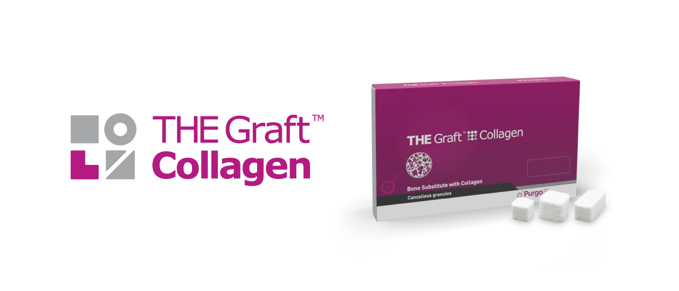
THE Graft Collagen
Last reviewed:
Answer-First Summary
THE Graft Collagen is a porcine-derived collagenated bone graft block (85% hydroxyapatite, 15% Type I collagen) for filling bone defects and bone augmentation. Although THE Graft Collagen is a medical device used in the dental field and is mentioned in relevant literature, the indications for the product must be determined based on the instructions for use and the expert's clinical opinion.
- Porcine-derived DPBM-C composition supports handling cohesion and scaffold function during healing.
- Osteoconductive properties and blood clot stability support bone formation during healing.
- Block and ring configurations for site-adapted trimming and placement.
- Documented uses in linked literature include alveolar ridge preservation, horizontal and/or vertical ridge augmentation, sinus floor augmentation, and regenerative periodontal treatment when aligned with labeling.
- Peer-reviewed items involving bovine collagenated comparators are cited with DOIs; interpret results from primary sources rather than assuming universal superiority.
Primary clinical use: space-maintaining graft and scaffold support for extraction socket and ridge contour preservation when consistent with approved indications.
Clinical Overview
THE Graft Collagen is used when clinicians need a shape-retentive xenograft that can be adapted to contained or semi-contained defects.
The block form is intended to act as a resorbable scaffold with handling properties that support contour control during implant placement, subject to clinical procedures and indications.
Size Selection Guide
| Size | Model | Recommended Use |
|---|---|---|
| 7x7x7 | TCB-01 | standard-sized extraction sockets |
| 8x9x10 | TCB-02 | moderate socket defects and ridge preservation procedures |
| 10x11x12 | TCB-03 | larger alveolar defects requiring volume augmentation |
| 3x5x7 | TCB-04 | narrow extraction sockets and localized periodontal defects |
| 5x5x5 | TCB-05 | narrow extraction sockets and localized periodontal defects |
| 5x5x10 | TCB-06 | standard-sized extraction sockets |
| 6x6x6 | TCB-07 | standard-sized extraction sockets |
| 10x20 | TCC-01 | bone defect filling and augmentation |
| 5x2.5 | TCHS-0105 | bone defect filling and augmentation |
| 5x2.5 | TCL-01 | bone defect filling and augmentation |
| 7x3x7 | TCL-02 | narrow extraction sockets and localized periodontal defects |
| 6x6 | TCO-01 | bone defect filling and augmentation |
| 5x11x18 | TCP-01 | moderate socket defects and ridge preservation procedures |
| 1.5x10x10 | TCP-02 | narrow extraction sockets and localized periodontal defects |
| 1.5x15x10 | TCP-03 | standard-sized extraction sockets |
| 5x3x8 | TCR-01 | narrow extraction sockets and localized periodontal defects |
| 5x4x9 | TCR-02 | narrow extraction sockets and localized periodontal defects |
| 5x5x10 | TCR-03 | standard-sized extraction sockets |
| 5 | TCS-01 | bone defect filling and augmentation |
| 12x11x10 | TCT-01 | larger alveolar defects requiring volume augmentation |
| 3x1x10 | TCW-01 | narrow extraction sockets and localized periodontal defects |
| 3x1x15 | TCW-02 | narrow extraction sockets and localized periodontal defects |
Clinical Selection Guide
THEGRAFTCOLLAGEN-INDEX is selected according to defect morphology and intended contour support, helping clinicians balance material efficiency with procedural predictability.
For manufacturer-reported hydration time under described test conditions, see the hydration time FAQ on this page (#faq).
Frequently Asked Questions
What are the sizes of THE Graft Collagen?
THE Graft Collagen is offered in multiple block and ring dimensions across the product line. For model numbers, dimensions (mm), and typical use contexts, see the Size Selection Guide on this page.
What kind of bone graft material is THE Graft Collagen?
THE Graft Collagen is a porcine-derived collagenated bone graft block (85% hydroxyapatite, 15% Type I collagen) for filling bone defects and bone augmentation. The DPBM-C composition (85:15) provides osteoconductive properties and blood clot stability for bone formation.
What are the components of THE Graft Collagen?
THE Graft Collagen is composed of 85% hydroxyapatite and 15% Type I collagen. The hydroxyapatite is derived from porcine (pig) cancellous bone, while the Type I collagen is also of porcine origin. The hydroxyapatite component provides the structural framework, while the collagen component enhances handling properties and stability. This composition creates a bone graft material with high purity and minimal presence of organic compounds that could potentially cause immunogenicity, as all organic compounds and lipids are removed through a manufacturer's virus inactivation process.
What is the shelf life of THE Graft Collagen?
The shelf life of THE Graft Collagen is 3 years from the date of manufacture.
What is the weight and volume ratio of bone graft material to collagen in THE Graft Collagen?
In THE Graft Collagen, the weight ratio of hydroxyapatite (bone mineral) to collagen is approximately 85:15. Due to freeze-drying, moisture from the collagen evaporates, which affects the apparent weight proportion compared with moisture-containing states. A reliable volume ratio is not specified in the available product documentation.
Is the collagen in THE Graft Collagen cross-linked?
The collagen in THE Graft Collagen is non-cross-linked collagen. It uses Type I collagen that has not undergone cross-linking processes.
What are the key material characteristics and clinical use considerations of THE Graft Collagen?
Key technical characteristics and clinical use considerations of THE Graft Collagen: 1. Source: THE Graft Collagen is derived from porcine bone mineral and porcine Type I collagen. 2. Structure: As a porcine-derived DPBM-C block and ring, THE Graft Collagen is manufactured to achieve a higher-density structure. This higher bone-graft content per unit volume can support space maintenance and volume stability during healing. - Space Maintenance/Volume Stability: Higher-density graft structures may provide longer-term contour support and space maintenance; clinical outcomes depend on defect configuration, technique, and patient factors. - Wettability/Handling: When hydrated with blood or physiological saline, THE Graft Collagen maintains cohesion, helping reduce particle scattering and making it easier to precisely trim and adapt to the desired form.
Is it necessary to use a membrane with THE Graft Collagen?
While THE Graft Collagen provides better handling properties than particulate bone grafts alone, it is generally recommended to use a membrane with it for optimal results, especially in non-contained defects. For optimal bone regeneration outcomes, particularly in guided bone regeneration (GBR) procedures, using THE Graft Collagen in combination with an appropriate membrane is recommended for better space maintenance and reduced graft resorption.
What are the advantages of using THE Graft Collagen for maxillary sinus augmentation?
The advantages of using THE Graft Collagen for maxillary sinus augmentation include optimized hydrophilic properties. THE Graft Collagen ensures reliable and uniform hydration, which may support procedural preparation efficiency during sinus lift procedures. ※ Results may vary depending on individual patient conditions
What are the differences between THE Graft and THE Graft Collagen?
THE Graft and THE Graft Collagen differ in composition and handling profile. Clinical application: THE Graft is used for filling various defects and requires a membrane for containment, while THE Graft Collagen is often selected for shaped defects and is easier to place and maintain position.
What are the indications for THE Graft Collagen?
Common indications described for THE Graft Collagen include: 1. Socket preservation and ridge preservation procedures - Particularly effective for maintaining alveolar ridge dimensions after tooth extraction - Available in block and ring types to fit different socket shapes and sizes 2. Intra-socket bone grafting - Used for filling extraction sockets while maintaining space 3. Ridge augmentation procedures - The moldable nature makes it ideal for adapting to ridge contours 4. Layering technique applications - Can be effectively combined with THE Graft in layered approaches 5. Periodontal intrabony defects - The collagen component helps with stability in these contained defects 6. Gap filling during immediate implant placement - Maintains position better than granular materials alone 7. Peri-implantitis dehiscence defects - The ring type is particularly useful for treating these defects THE Graft Collagen is often discussed for contained defects where its handling properties, moldability, and volume maintenance capabilities provide significant advantages over traditional granular materials. ※ Results may vary depending on individual patient conditions
Are there any tips for using THE Graft Collagen?
Tips for using THE Graft Collagen: 1. Hydration technique: - Can be used in original condition or hydrated with saline or blood - Molding is often easier after hydration 2. Shaping and adaptation: - Use sterile scissors or blades to trim the block to the desired shape before placement - Avoid excessive pressure when molding to preserve the porous framework 3. Layering approach: - Layering approaches vary by defect morphology and clinician preference; if combining particulate graft and a collagenated block/ring, adapt materials to maintain space and contour, following IFU and case-specific planning 4. Membrane coverage: - Cover with an appropriate barrier membrane when indicated - Both resorbable and non-resorbable membranes may be used depending on the case 5. Placement considerations: - Avoid overfilling the defect site - Ensure good adaptation to defect walls for stable positioning 6. Storage: - Store at temperatures between 15°C-25°C (59°F-77°F) - Keep away from direct sunlight 7. Handling: - Always use sterile surgical instruments and gloves when handling the material - THE Graft Collagen is for single use only - never reuse or re-sterilize
How much volume reduction occurs as the collagen dissolves in THE Graft Collagen? What is the percentage of volume reduction?
Based on the cited summaries, reported dimensional or volumetric changes may occur over time as healing and remodeling progress. The collagen component (described in some materials as approximately 14-17% of the initial volume) is expected to resorb over time. Treatment planning should follow IFU indications and clinician judgment; outcomes vary by defect type, technique, and patient factors. Paper: "Particulate Versus Cross-Linked Collagenated Bone Substitutes for Guided Bone Regeneration: A Randomized Controlled Trial" (see listed references for DOI and details). ※ Results may vary depending on individual patient conditions
Why is THE Graft Collagen difficult to see on X-rays?
THE Graft Collagen is difficult to visualize on X-rays due to its high porosity. While this makes it less visible radiographically initially, the porous structure creates space for bone cell attachment and new bone formation under stated conditions. As healing progresses, the grafted area may gradually show increased radiopacity on follow-up imaging.
Should THE Graft Collagen be molded when using it? How much pressure should be applied during molding?
THE Graft Collagen may be molded and adapted to fit the defect morphology. Regarding pressure: available documentation does not specify a single numeric pressure value. In practice, clinicians typically use light manual pressure to adapt the block/ring while avoiding excessive compression that could collapse the porous framework. Final handling technique should follow IFU guidance, defect configuration, and clinician judgment.
Should THE Graft Collagen be hydrated with saline or blood?
Moistening with saline makes it easier to cut into pieces and shape appropriately. THE Graft Collagen can also be hydrated with blood per IFU and case preference.
What is the appropriate hydration time for THE Graft Collagen?
THE Graft Collagen has a reported short hydration time. Manufacturer and study summaries for this line report full hydration within seconds under described test conditions; clinical chairside may vary on the procedure and model.
What is the remodeling timeline for THE Graft Collagen?
THE Graft Collagen shows the following remodeling timeline in cited summaries: Significant bone formation is described as beginning at around 4-5 months, with further maturation commonly discussed at later follow-up (e.g., 5-6 months). Remodeling is generally discussed over approximately 1 year, with variation depending on defect type, technique, and patient factors. Some cited summaries describe cortical line formation becoming visible within the first year. In histological analyses at various time points, new bone formation surrounding residual graft material has been described. Treatment planning and timing should follow IFU indications and clinician judgment. ※ Results may vary depending on individual patient conditions
Are there any research papers related to THE Graft Collagen?
Peer-reviewed studies involving THE Graft Collagen are listed with full citations and DOIs in the Clinical Evidence and Reference (Academic Evidence) sections on this page (#clinical-evidence).
Does the presence of collagen in bone graft materials affect bone remodeling?
Effect on bone quality: Although there are concerns that a large amount of collagen in bone grafts may degrade bone quality, bone graft materials containing an appropriate amount of collagen help maintain stable volume and promote vascularization during bone formation. Effects on remodeling process: Collagen in bone blocks can enhance guided bone regeneration. Collagen stabilizes the blood clot, maintaining a high concentration of autogenous growth factors. Collagen promotes osteoconductivity, increasing the percentage of new bone formation and reducing residual grafting material. Practical handling characteristics discussed in clinical reports despite potential quality concerns: Collagen-containing materials offer better handling properties and stability. They show high hydrophilicity due to their high surface area and porosity. They promote hemostasis and angiogenesis, and induce cell migration necessary for regeneration. ※ Results may vary depending on individual patient conditions
Can THE Graft Collagen be used with screws or pins?
Yes, THE Graft Collagen can be used with screws and pins: 1. Clinical cases demonstrate the combined use of THE Graft Collagen with: - Bone tacks for stabilization in GBR procedures - Tenting screws in guided bone regeneration techniques 2. The combination provides several benefits: - Improved stability of the graft material during healing - Better space maintenance for bone regeneration - Enhanced predictability of the bone augmentation procedure 3.Specific techniques documented in clinical cases: - "Tenting screw with implant simulation" technique using THE Graft Collagen - Immediate implant placement with bone graft stabilized by tacks 4. Surgical protocols often include: - Placement of tenting screws or pins first - Application of THE Graft Collagen around the hardware - Coverage with a resorbable or non-resorbable membrane - Stabilization with additional tacks if needed This combination approach helps maintain the volume of the augmented site and prevents collapse of the overlying membrane, leading to more predictable bone regeneration outcomes. ※ Results may vary depending on individual patient conditions
How do you trim THE Graft Collagen blocks?
THE Graft Collagen blocks can be trimmed with sterile surgical scissors or a scalpel to the desired shape. Trim while dry for more precise cutting, or hydrate first in saline for 1-2 minutes for easier shaping. Shape the block to match defect contours; for irregular defects, smaller pieces may be packed together. After placement, the trimmed block can be further adapted in situ using light pressure.
How is THE Graft Collagen Ring Type used?
THE Graft Collagen Ring Type is offered in sizes including 5x3x8mm, 5x4x9mm, and 5x5x10mm (Height x Inner Ø x Outer Ø). It is used for socket preservation, ridge preservation, and peri-implantitis defects by placing the ring into the extraction socket, over a ridge defect, or around an exposed implant surface to maintain space during healing.
Why is collagen added to bone graft materials?
Collagen is added to bone graft materials for the following reasons: 1. Improved handling properties: - Enhances manipulation and placement during surgery - Allows the material to be easily shaped to fit bone defects - Provides better stability and pressure transmission 2. Biological benefits: -Promotes hemostasis and angiogenesis due to high hydrophilicity - Induces cell migration necessary for regeneration - Stabilizes blood clots, maintaining high concentration of autogenous growth factors - Promotes osteoconductivity, increasing percentage of new bone formation 3. Structural advantages: - Creates a composite material that mimics natural bone matrix - Maintains space for blood vessel formation through trabeculae structure - Provides resistance to degradation while allowing controlled resorption 4. Clinical performance: - Increases bone quantity in sites with insufficient bone for implants - Induces bone formation in defect areas - Facilitates handling during surgery while maintaining volume after surgery - Collagen-containing bone grafts are described as offering handling advantages in terms of manipulation and stability, making them particularly useful for bone augmentation procedures.
What type of collagen (Type I, Type III, etc.) is used in bone graft materials?
Type I collagen is used in THE Graft Collagen bone graft material. Specifically: 1. Source: - THE Graft Collagen incorporates Type I collagen derived from porcine tendon 2. Verification methods: - The Type I collagen is verified through SDS-PAGE analysis - Amino acid analysis confirms it as Type I collagen by detecting: - High mol% of hydroxyproline - Minimal amounts of tyrosine, tryptophan, and cysteine 3. Quality control: - Amino acid analysis is performed for every lot of THE Graft Collagen - This ensures consistency and high quality by verifying the collagen type 4. Safety features: - The Type I collagen undergoes virus inactivation processes - It is processed to remove terminal telopeptides that could induce antigenicity and immunogenicity - The collagen is processed into atelocollagen form by enzyme treatment of N-terminal and C-terminal regions Type I collagen is specifically chosen because it is the predominant collagen type in bone tissue, providing structural support while maintaining biocompatibility.
What is the source of collagen (porcine, bovine, synthetic, etc.) used in bone graft materials?
The collagen in THE Graft Collagen is derived from porcine (pig) sources. Specifically: 1. Source details: - The Type I collagen is extracted from porcine tendon - The porcine raw materials originate from Korea and Australia 2. Advantages of porcine-derived collagen: - Porcine bones and tendons have a relatively low risk of zoonosis compared to bovine sources - Porcine-derived materials are free from the risk of Creutzfeldt-Jakob Disease - The crystalline and morphological structures of porcine bone resemble human cancellous bone - Porcine bone's porosity structure and form are more similar to human bone, providing better biocompatibility 3. Safety considerations: - Porcine materials, unlike bovine bone and cattle ligaments, don't require management of TSE (Transmissible Spongiform Encephalopathy) agents - Raw materials undergo safe practices adhering to regulatory standards (HACCP, GMP) - The collagen is processed through virus inactivation to eliminate infection risk Regulatory status: - THE Graft Collagen with porcine-derived collagen is approved for use in 10 countries including Korea and EU nations - It was the first application of MDR (Medical Device Regulation) with THE Graft in Korea as a xenograft for EU The choice of porcine sources represents a strategic decision to maximize biocompatibility while minimizing potential risks associated with other animal-derived materials.
Is THE Graft Collagen registered in the Korean Health Insurance Review and Assessment Service (HIRA) insurance system?
THE Graft Collagen may be listed as a biomaterial for tissue repair or bone graft material under Korean national health insurance when use aligns with approved indications and current HIRA/EDI schedules. Whether a specific case is reimbursable depends on the diagnosis, procedure, and billing rules at the time of treatment. In accordance with HIRA standards, reimbursement is generally considered only when use matches approved diagnostic codes, such as bone defects related to osseous lesions or certain periodontal regenerative indications. Bone grafting performed solely for routine implant placement is often classified as non-reimbursable. Clinics should confirm the applicable EDI code and listing status for the specific THE Graft Collagen model in the hospital billing system (OCS/EMR) against the latest official schedules. Use should follow the instructions for use and locally approved labeling.
Is THE Graft Collagen widely used internationally?
THE Graft Collagen is used internationally in multiple markets: 1. Market presence: - THE Graft Collagen is sold in 10 countries including Korea and European Union member states 2. Regulatory achievements: - The product has achieved MDR (Medical Device Regulation) approval for the European market - It was noted as "the first application of MDR with THE Graft in Korea as a xenograft (EU)" 3. International clinical applications: - Clinical cases and studies from different countries demonstrate its use in various dental procedures including: - Socket preservation - Ridge preservation - Peri-implantitis dehiscence defect treatment While the search results confirm international usage across multiple countries, they don't provide specific market share data or comparative usage statistics between countries. The regulatory approval in the EU market suggests compliance with international standards and acceptance in developed healthcare markets. Use should follow the instructions for use and locally approved labeling.
Is THE Graft Collagen being used in dental university hospitals in Korea?
THE Graft Collagen is used in multiple Korean dental university hospitals and has been featured in various academic research papers. (*This information does not guarantee the efficacy or effects of the product. Specific use should follow the instructions for use and the clinical judgment of a qualified professional.)
What material factors can influence perceived firmness of THE Graft Collagen during handling?
THE Graft Collagen is manufactured using optimized process, resulting in a high density and tightly combined bone graft material; consequently, it offers volume retention and maximizes clinical predictability.
What is the reason for developing the Ring Type of THE Graft Collagen?
the Ring Type of THE Graft Collagen appears to have been developed for specific clinical applications: 1. Implant placement: - The Ring Type is designed to facilitate simultaneous implant placement and bone grafting - It allows for immediate implant placement while providing bone augmentation around the implant 2. Defect-specific design: - The Ring Type is particularly useful for treating specific defect configurations - It provides better adaptation to certain types of bone defects, especially those with vertical dimension needs 3. Available in multiple sizes: - TCR-01: 5x3x8 mm - TCR-02: 5x4x9 mm - TCR-03: 5x5x10 mm - These varying dimensions allow for selection based on the specific defect size and implant diameter 4. Clinical applications: - Used in ridge preservation procedures - Particularly effective in cases with buccal bone deficiency - Provides a structural framework for bone regeneration while allowing implant placement The Ring Type appears to be designed to address the specific clinical need for simultaneous implant placement and bone augmentation, offering a convenient solution that maintains space and provides structural support during healing.
Is THE Graft Collagen suitable for socket preservation? How should it be properly used for this application?
Yes, THE Graft Collagen is suitable for socket preservation procedures. here's how it can be properly used: Application Methods: 1. Single Use Method: - THE Graft Collagen can be used alone to fill the extraction socket - The material should be compactly filled into the extraction socket - After filling, cover with a resorbable collagen membrane - Secure with interrupted and horizontal mattress sutures (4-0 e-PTFE Biotex® recommended) 2. Layering Technique: - Place THE Graft (particulate form) at the bottom of the socket - Place THE Graft Collagen on top - This combination technique may provide better bone quality according to some clinicians Histological Analysis: - Histological evaluation 6 months after socket preservation showed residual bone material surrounded by new bone, indicating successful biochemical fusion Benefits for Socket Preservation: - High porosity promotes effective new bone formation - Favorable packing ability and pressure transmission - Hydrates easily for precise control over graft material - High stability and bone regeneration efficiency THE Graft Collagen is particularly useful for socket preservation due to its handling properties and ability to maintain the extraction socket volume while promoting bone regeneration.
Is THE Graft Collagen suitable for alveolar ridge preservation? How should it be properly used for this application?
Yes, THE Graft Collagen is suitable for alveolar ridge preservation. here's how it can be properly used: Application Methods: 1. Standard Technique: - Compactly fill THE Graft Collagen into the extraction socket - Cover with a resorbable collagen membrane - Secure with interrupted and horizontal mattress sutures 2. L-Technique: - Shape THE Graft Collagen in an L-configuration to match the defect - Apply membrane over the graft material - Fix the membrane using sutures - This technique is particularly useful for ridge preservation in posterior areas 3. Layering Technique: - Place THE Graft (particulate form) as the base layer - Add THE Graft Collagen (ring type) on top - Use an open barrier membrane technique - This combination may provide better bone quality according to clinical experience Clinical Case Examples: Effectiveness: - THE Graft Collagen has been used successfully in both intact and damaged extraction sockets - Histological analysis shows residual bone material surrounded by new bone, indicating biochemical fusion - Studies demonstrate effective ridge preservation with minimal dimensional changes THE Graft Collagen is particularly effective for ridge preservation due to its handling properties, volume stability, and ability to promote bone regeneration while maintaining the alveolar ridge dimensions. Use should follow the instructions for use and locally approved labeling.
When using THE Graft Collagen for socket preservation, is it necessary to use a membrane on top?
Although THE Graft Collagen provides better handling properties than particulate bone grafts alone, the use of a membrane is generally recommended for optimal results. 1. Documented cases: A resorbable collagen membrane is used in conjunction with THE Graft Collagen for socket preservation. 2. Clinical evidence: In non-contained defects, using THE Graft Collagen without a membrane may lead to greater ridge dimension loss, so membrane coverage is recommended. 3. Membrane functions: - Maintains space and stabilizes the graft material. - Prevents the graft from dispersing. - Enhances bone regeneration outcomes. 4. Intact periosteum considerations: - While some literature suggests intact periosteum may provide similar effects to resorbable membranes, clinical experience indicates that periosteum alone cannot provide the same space maintenance and graft stabilization that membranes offer. 5. Exception: - If the bone defect has a shape that naturally prevents graft material dispersion, membrane use might be less critical. In conclusion, while THE Graft Collagen has favorable handling characteristics compared to particulate grafts, using a membrane is still recommended for optimal outcomes in socket preservation, particularly in non-contained defects where graft stability is needed. Use should follow the instructions for use and locally approved labeling.
Is THE Graft Collagen suitable for horizontal and vertical bone augmentation? How should it be properly used for these applications?
Yes, THE Graft Collagen is suitable for both horizontal and vertical bone augmentation procedures. Here's how it should be properly used: For Horizontal Bone Augmentation: 1. Application Methods: - Use THE Graft Collagen in block form - Trim the block to an L-shape to fit the defect area - Cover with a collagen membrane , a resorbable collagen membrane or THE Cover) - Secure primary closure using retentive flap technique 2. Clinical Outcomes: - THE Graft Collagen provides good handling properties and stability for horizontal augmentation For Vertical Bone Augmentation: 1. Application Methods: - Mix with autogenous bone when possible for better outcomes - Use titanium-reinforced membranes (like OpenTex-TR) or titanium mesh for space maintenance - Consider using THE Graft Collagen with THE Graft particulate for better stability 2. Important Considerations: - Vertical augmentation is most predictable up to 3mm from the host bone - When using only bone substitutes without autogenous bone, longer healing periods (10+ months) are recommended - For vertical augmentation beyond 3mm, consider mixing with autogenous bone or growth factors , rhBMP-2) - Adequate space maintenance using titanium-reinforced membranes is crucial THE Graft Collagen offers advantages for both horizontal and vertical bone augmentation, including improved handling, shape stability, and space maintenance compared to particulate grafts alone. However, technique-sensitive procedures like vertical augmentation require careful planning and execution for predictable outcomes.
Is THE Graft Collagen suitable for maxillary sinus bone grafting? How should it be properly used for this application?
Yes, THE Graft Collagen is suitable for maxillary sinus bone grafting procedures. here's how it can be properly used: Application Methods: 1. Lateral Window Approach: - Create a lateral window to access the sinus cavity - Carefully elevate the sinus membrane - Apply THE Graft Collagen (collagenated block bone substitute) to the elevated sinus - Place a resorbable collagen membrane over the lateral window - Suture the site appropriately 2. Crestal Approach: - Can be used with osteotome sinus floor elevation techniques - Both simple techniques and bone-added osteotome sinus floor elevation (BAOSFE) techniques are viable - In cases with residual bone height of 5mm or less, bone grafting is recommended 3. Implant Placement Timing: - Can be used for both simultaneous implant placement and staged approaches - For simultaneous placement, ensure primary stability of the implant Clinical Evidence: - Studies show successful bone formation and osseointegration when using THE Graft Collagen in sinus augmentation procedures - Comparative studies with bovine-derived materials show similar clinical outcomes - THE Graft Collagen has been used successfully in various sinus augmentation techniques, including lateral window and crestal approaches Advantages for Sinus Grafting: - The collagen component provides better handling properties - Maintains volume stability in the sinus cavity - Can be easily shaped to fit the sinus cavity - Hydrates easily for precise control over the amount of graft material - High stability and bone regeneration efficiency THE Graft Collagen is an effective option for maxillary sinus bone grafting procedures, offering good handling properties, volume stability, and successful clinical outcomes when used according to proper surgical protocols. Use should follow the instructions for use and locally approved labeling.
Does THE Graft Collagen maintain good volume and achieve sufficient ossification even in 1-wall defects after 3-4 months?
Based on clinical studies, THE Graft Collagen shows limited effectiveness in 1-wall defects compared to 2-wall and 3-wall defects. Volume stability was poorer compared to multi-wall defects due to fewer surrounding walls providing support for the graft material. Therefore, if guided properly using a membrane, sufficient volume is maintained even in a 1-wall defect, and ossification occurs effectively. Use should follow the instructions for use and locally approved labeling.
Will bone created from collagen-type bone graft materials maintain well even after 10-20 years?
The collagen contained in THE Graft Collagen is completely absorbed within one month after implantation. Thereafter, the remaining THE Graft acts as a scaffold, influencing new bone formation and bone remodeling. Bone remodeling generally takes about 1 year, with significant variation depending on graft material type. THE Graft shows reported with shorter timing in some cited summaries remodeling, with cortical line formation visible within 1 year, which may indicate earlier new bone formation in cited summaries. For long-term stability specifically, the available data suggests xenograft materials persist in the body longer than autografts or allografts, providing structural support while natural bone forms around them.
For extensive bone defects such as 1-wall defects, is it better to combine THE Graft Collagen with a resorbable membrane or a non-resorbable membrane?
Non-resorbable membranes are generally preferred for extensive defects like 1-wall defects because they provide better space maintenance and structural support When using THE Graft Collagen in wide defect areas, non-resorbable membranes or mesh are recommended to prevent membrane exposure and ensure better outcomes Clinical experiences indicate that non-resorbable membranes are more effective for horizontal bone augmentation in large defects, though they carry a risk of exposure For cases with significant buccal bone recession, collagen membranes are recommended over collagen matrix materials to provide better stability for the bone graft
When should this size be selected for clinical use?
THE Graft Collagen Product Line Overview in this size is recommended for dental bone defect management in guided regenerative workflows. This size-focused allocation helps reduce material waste and supports precise graft adaptation to the defect boundary.
Why can this block type be preferred over particulate grafts?
Compared with particulate grafts, this collagenated block format is designed for improved handling cohesion and space maintenance at the recipient site. For reported hydration time under described test conditions, see the hydration time FAQ on this page (#faq).
Clinical Evidence
Clinical evidence for THE Graft Collagen is listed in citation format with journal, year, and DOI. Comparative outcomes and source tables are detailed in the Reference (Academic Evidence) section below.
Jae-Hong Lee, Eun-Hee Jung, Seong-Nyum Jeong. Profilometric, volumetric, and esthetic analysis of guided bone regeneration with L-shaped collagenated bone substitute and connective tissue graft in the maxillary esthetic zone: A case series with 1-year observational study. Clinical Implant Dentistry and Related Research 2022. DOI: https://doi.org/10.1111/cid.13116 [Ref 1]
Jae-Hong Lee, Eun-Hee Jung, Seong-Nyum Jeong. Augmentation Stability of Guided Bone Regeneration for Peri-Implant Dehiscence Defects with L-shaped Porcine-Derived Block Bone Substitute. Materials 2021. DOI: https://doi.org/10.3390/ma14216580 [Ref 2]
Jae-Hong Lee, Seong-Nyum Jeong. Use of Porcine-derived Block Bone Substitutes for Guided Bone Regeneration in the Peri-implant Dehiscence Defects of the Mandibular Anterior Region. Journal of implantology and applied sciences 2021. DOI: https://doi.org/10.32542/implantology.2021018 [Ref 3]
Jae-Hong Lee, Ji-Hoo Han, Seong-Nyum Jeong. Porcine-derived soft block bone substitutes for the treatment of severe class II furcation-involved mandibular molars: a prospective controlled follow-up study. Journal of Periodontal & Implant Science 2022. DOI: https://doi.org/10.5051/jpis.2203660183 [Ref 4]
Jae-Hong Lee, Yeon-Tae Kim, Seong-Nyum Jeong. Alveolar ridge preservation of damaged or periodontally compromised extraction sockets with bovine- and porcine-derived block bone substitutes: A retrospective case–control study. Clinical Implant Dentistry and Related Research 2023. DOI: https://doi.org/10.1111/cid.13246 [Ref 5]
Yeon-Tae Kim, Jae-Hong Lee. Profilometric, esthetic, and patient-reported outcomes of the L-shape technique combined with delayed connective tissue grafting in the maxillary anterior region: A 3-year case series study. Journal of Esthetic and Restorative Dentistry 2024. DOI: https://doi.org/10.1111/jerd.13229 [Ref 6]
Ji-Young Jung, Seung-Hyun Park, Kwan-Jung Kim, Kyung-A Ko, Dong-Woon Lee, Jung-Seok Lee. Particulate Versus Cross-Linked Collagenated Bone Substitutes for Guided Bone Regeneration: A Randomized Controlled Trial. Clinical Oral Implants Research 2025. DOI: https://doi.org/10.1111/clr.14433 [Ref 7]
Ji-Young Yoon, Jin-Won Choi, Pil-Young Yun, Jeong-Kui Ku. Clinical and radiographic evaluation of bone graft after apicoectomy using porcine-derived collagenous block type xenograft. Journal of Dental Implant Research 2025. DOI: https://doi.org/10.54527/jdir.2025.44.1.8 [Ref 8]
Jin-Won Choi, Soo-Shin Hwang, Pil-Young Yun, and Jeong-Kui Ku. Mesiodistal Marginal Bone Loss around Implants with Porcine Xenografts: A Preliminary Retrospective Comparison of Collagenous Block and Powder Forms. Journal of Hard Tissue Biology 2025. DOI: https://doi.org/10.2485/jhtb.34.205 [Ref 9]
Dongseob Lee, Ki-Tae Koo, Yang-Jo Seol, Yong-Moo Lee, Jungwon Lee. Comparison of osteogenesis and osseointegration following implant placement with simultaneous maxillary sinus augmentation using particulate and collagenated block types of deproteinized porcine bone mineral: a radiographic and histomorphometric analysis. Clinical Oral Investigations 2023. DOI: https://doi.org/10.1007/s00784-023-05197-7 [Ref 10]
Dongseob Lee, Jin Uk Choi, Young-Chang Ko, Ki-Tae Koo, Yang-Jo Seol, Yong-Moo Lee, Jungwon Lee. Does defect configuration affect the outcomes of alveolar ridge preservation? An experimental in vivo study. Journal of Periodontal & Implant Science 2024. DOI: https://doi.org/10.5051/jpis.2401480074 [Ref 11]
Instruction for Use
Instruction for Use (EN_IFU)
DESCRIPTION
THE Graft Collagen, a form of block and ring composed of porcine derived bone mineral matrix from cancellous bone and Type I Collagen from porcine tendon, is a material used to fill, augment, and/or reconstruct periodontal, oral, and maxillofacial defects. The bone mineral matrix is similar to physical and chemical aspects of the mineralized matrix of human bone. THE Graft Collagen can be trimmed in various shapes depending on the bone defect. THE Graft Collagen is sterilized using gamma irradiation and recommended for the adult* patient who needs filling of bone defects and bone augmentation. In order to place dental implants in THE Graft Collagen augmented area, based on clinical experience, it takes several months to gain bone stability through THE Graft Collagen's partial transformation by the osteoclast and osteoblast.
* In accordance with the adult standards applicable in each country.
INDICATIONS
THE Graft Collagen is recommended for the patient who needs filling of bone defects and bone augmentation e.g.:
- Alveolar ridge preservation
- Horizontal and/or vertical ridge augmentation
- Sinus floor augmentation
- Regenerative periodontal treatment
DIRECTIONS FOR USE
Preparation before Use
- Check Use by Date and condition of the package of the product.
- Use the device in a dental hospital and dental clinics.
INSTRUCTIONS
- After exposure of the bony defect with a mucoperiosteal flap, all granulation tissue must be carefully removed when it needs.
- In order to assure the formation of new bone, THE Graft Collagen should only be placed in direct contact with well-vascularized bone.
- The mucoperiosteal flaps should be sutured to achieve primary closure, if possible. A surgical dressing may be placed over the wound for one to two weeks.
- If primary wound closure cannot be achieved completely, further immobilization of the flap (e.g., by incision through the periosteum) should be performed and/or a bioabsorbable membrane should be placed over the bone graft site.
Procedure
- Using sterile equipment and/or sterile gloves, remove the product from the package.
- Check the surgical site(s).
- If necessary, size the bone block to desired shape and/or form using sterilized scissors and/or blades.
- THE Graft Collagen can be used in its original condition. THE Graft Collagen can also be hydrated with normal saline solution [0.9% w/v of Sodium Chloride] or patient's blood.
- Implant the product at the site of bone defect.
- Avoid excessive pressure on particles or overfilling the defect site.
- After implant cover up surrounding tissues using membrane. Both Resorbable and non-resorbable membrane is available but Purgo's membrane such as THE Cover, OpenTex or OpenTex-TR is strongly recommended.
Pre-Operative care
- It should be determined whether the patient has any concerning medical conditions (e.g., diabetes, osteoporosis, allergies, autoimmune disease, etc.), is currently taking any medications (e.g., steroids, aspirin, anticoagulants, bisphosphonate, etc.), and their smoking status.
- Before bone grafting, it is imperative to undergo treatment for periodontal disease and adhere to oral hygiene measures. This helps in minimizing potential complications associated with the surgery.
Post-Operative care
- The surgeon should instruct patients not to apply pressure to the surgical site during the bone healing process.
- The surgeon should inspect and evaluate patients for any allergic response due to the grafted device.
- The grafted site should be allowed to heal for at least 3 to 6 months prior to implant placement.
- Successful periodontal treatment requires adequate oral hygiene and infection control.
- Removal of the grafted device is seldom required. However, in case of severe systemic infection or traumatic injury, early removal may be necessary.
CONTRAINDICATIONS
THE Graft Collagen should not be used in patients with the following conditions:
- Known allergies to collagen,
- Uncontrolled periodontal diseases,
- Uncontrolled metabolic and endocrine disorders that affect bone health (e.g. diabetes, osteoporosis, osteomalacia, primary hyperparathyroidism etc.),
- Undergoing radiotherapy, chemotherapy, or immunosuppressive therapy,
- During pregnancy or breastfeeding,
- Heavy smoking.
PRECAUTIONS/WARNINGS
1. Do not use if package has been opened or damaged prior to use.
2. Do not reuse or re-sterilize THE Graft Collagen. Inflammation, fever, edema (swelling) may occur upon re-use.
3. THE Graft Collagen has been designed for single use only. Reusing this medical device bears the risk of cross-patient contamination.
4. The Graft Collagen is not designed for use under load bearing conditions. Mechanical loading (compression loading) of THE Graft Collagen augmented areas is possible after 4-6 months at the earliest (appropriate healing time can be adjusted according to the defect size and wound healing).
5. THE Graft Collagen should be kept in sterile condition until using the product.
6. THE Graft Collagen must be handled by qualified professionals, trained dentists, and/or oral surgeons.
7. It is not recommended to combine with other bone graft materials; however, it can be used with other materials according to the expert's clinical opinion.
8. THE Graft Collagen has not been evaluated for safety and compatibility in the MR environment. It has not been tested for heating, migration, or image artifact in the MR environment.
SIDE EFFECTS
Possible side effects may occur during the healing period, including but not limited to:
1. Initial healing phase
- Hematoma, infection, wound dehiscence, membrane exposure, overlying tissue necrosis, facial swelling, and contusion.
2. Late healing phase
- Severe volumetric collapse, implant failure related to the grafted site.
It should be noted that the aforementioned complications can also be caused by surgical errors and should be interpreted with caution.
STORAGE AND HANDLING
1. THE Graft Collagen should be stored at the temperature between 15℃ - 25℃ (59°F – 77°F) in moderate humidity condition range from 0 to 70%.
2. THE Graft Collagen should be kept away from direct sunlight.
3. THE Graft Collagen should be handled with sterile surgical instruments and/or gloves.
DISPOSAL
- Once THE Graft Collagen is removed from its packaging, the product must be properly discarded, even when the product is not used.
- THE Graft Collagen is disposed of according to the applicable country's medical device disposal method.
AVAILABILITY
- THE Graft Collagen is available in various sizes and forms. Please visit our website for more information.
SYMBOLS GLOSSARY
Do not reuse Catalogue number
Sterilization by irradiation Cause
Do not resterilize Use by date
Manufacturer CE mark and identification number of notified body
Authorized representative in the European Community Batch code
Temperature limitation Do Not Use if package is damaged and consult instructions for use
Keep away from sunlight Caution: US Federal law restricts this device to sale by or on the order of a licensed dentist (For US Only)
Non-pyrogenic Consult instructions for use or consult electronic instruction for use
Contains biological material of animal origin
Country of manufacture Unique device identifier
Medical device Single sterile barrier system with protective packaging inside
Date of manufacture Use-by date
Keep dry Importer
Patient name Patient information website
Patient identification Health care center or doctor
Date Humidity Limitation
When a serious incident has occurred during use of THE Graft Collagen, it should be reported to the manufacture and the competent authority where the user and/or patient is established.
Instructions for use into other languages refer to the following site. www.purgo-pdr.com
THE Graft Collagen is a mixture of 85 wt-% THE Graft (over 95% hydroxyapatite) and 15 wt-%(±10 wt-%) porcine collagen fibers in block and ring form.
Manufacturer:
Purgo Biologics Inc.
#812 27 Dunchon-daero 457beon-gil, Jungwon-gu, Seongnam-si, Gyeonggi-do, Korea
Made in Korea
purgobio.com
Reference (Academic Evidence)
Clinical Implant Dentistry and Related Research
Jae-Hong Lee, Eun-Hee Jung, Seong-Nyum Jeong. Profilometric, volumetric, and esthetic analysis of guided bone regeneration with L-shaped collagenated bone substitute and connective tissue graft in the maxillary esthetic zone: A case series with 1-year observational study. Clinical Implant Dentistry and Related Research 2022. DOI: https://doi.org/10.1111/cid.13116
Ref 1Jae-Hong Lee, Yeon-Tae Kim, Seong-Nyum Jeong. Alveolar ridge preservation of damaged or periodontally compromised extraction sockets with bovine- and porcine-derived block bone substitutes: A retrospective case–control study. Clinical Implant Dentistry and Related Research 2023. DOI: https://doi.org/10.1111/cid.13246
Ref 5Control group: Bio-Oss Collagen
Comparative performance: For the horizontal width, DPBM-C group showed a decrease of 1.66 ± 1.80 mm (20.82%), while the DBBM-C group decreased by 1.70 ± 2.26 mm (21.50%) For the vertical height, DPBM-C group decreased by 1.44 ± 1.97 mm (27.89%), compared to 1.39 ± 1.85 mm (30.47%) in the DBBM-C group. For the buccal tissue contour, DPBM-C group showed a decrease of 2.81 ± 1.93 mm, while the DBBM-C group showed a decrease of 3.07 ± 1.44 mm
Source: Table 2 (p. 7)
Materials
Jae-Hong Lee, Eun-Hee Jung, Seong-Nyum Jeong. Augmentation Stability of Guided Bone Regeneration for Peri-Implant Dehiscence Defects with L-shaped Porcine-Derived Block Bone Substitute. Materials 2021. DOI: https://doi.org/10.3390/ma14216580
Ref 2Control group: Bio-Oss Collagen
Comparative performance: Implant Stability (Periotest Values - PTV): This was the parameter with a statistically significant difference (p = 0.042) during the re-entry surgery. DPBM-C Group: -4.52 ± 1.16 DBBM-C Group: -5.19 ± 1.22
Source: Figure 5(p.8)
Journal of implantology and applied sciences
Jae-Hong Lee, Seong-Nyum Jeong. Use of Porcine-derived Block Bone Substitutes for Guided Bone Regeneration in the Peri-implant Dehiscence Defects of the Mandibular Anterior Region. Journal of implantology and applied sciences 2021. DOI: https://doi.org/10.32542/implantology.2021018
Ref 3Journal of Periodontal & Implant Science
Jae-Hong Lee, Ji-Hoo Han, Seong-Nyum Jeong. Porcine-derived soft block bone substitutes for the treatment of severe class II furcation-involved mandibular molars: a prospective controlled follow-up study. Journal of Periodontal & Implant Science 2022. DOI: https://doi.org/10.5051/jpis.2203660183
Ref 4Control group: THE Graft
Comparative performance: Probing Pocket Depth (PPD) Reduction:DPBM-C: 4.1 ± 3.0 mm. Clinical Attachment Level (CAL) Gain:DPBM-C: 4.4 ± 2.9 mm. Vertical Furcation Defect (VFD) Reduction:DPBM-C: 4.1 ± 2.5 mm. The researchers noted that although the results were statistically similar, the DPBM-C group consistently showed higher values of improvementThis is likely because the soft block bone substitute provides better space maintenance and mechanical support in severe furcation defects compared to the particulate.
Source: Table2,Figure2(P.6)
Dongseob Lee, Jin Uk Choi, Young-Chang Ko, Ki-Tae Koo, Yang-Jo Seol, Yong-Moo Lee, Jungwon Lee. Does defect configuration affect the outcomes of alveolar ridge preservation? An experimental in vivo study. Journal of Periodontal & Implant Science 2024. DOI: https://doi.org/10.5051/jpis.2401480074
Ref 11Control group: Spontaneous healing
Comparative performance: The 3-wall defect exhibited a significantly higher percentage of mineralization in the ARP group after 12 weeks of healing (ARP: 61.73%±7.52%; SH: 48.84%±3.06%; P=0.029).
Source: Table 2(p. 12)
Journal of Esthetic and Restorative Dentistry
Yeon-Tae Kim, Jae-Hong Lee. Profilometric, esthetic, and patient-reported outcomes of the L-shape technique combined with delayed connective tissue grafting in the maxillary anterior region: A 3-year case series study. Journal of Esthetic and Restorative Dentistry 2024. DOI: https://doi.org/10.1111/jerd.13229
Ref 6Clinical Oral Implants Research
Ji-Young Jung, Seung-Hyun Park, Kwan-Jung Kim, Kyung-A Ko, Dong-Woon Lee, Jung-Seok Lee. Particulate Versus Cross-Linked Collagenated Bone Substitutes for Guided Bone Regeneration: A Randomized Controlled Trial. Clinical Oral Implants Research 2025. DOI: https://doi.org/10.1111/clr.14433
Ref 7Control group: THE Graft
Comparative performance: The collagenated group exhibited higher hard tissue dimensions in terms of horizontal width and augmented area. HT0 values were significantly higher in the collagenated group compared to the particulate group immediately after the surgery (3.64 [1.07] mm and 2.79 [1.02] mm, respectively; p = 0.0021).
Source: Table 1 (p. 7)
Journal of Dental Implant Research
Ji-Young Yoon, Jin-Won Choi, Pil-Young Yun, Jeong-Kui Ku. Clinical and radiographic evaluation of bone graft after apicoectomy using porcine-derived collagenous block type xenograft. Journal of Dental Implant Research 2025. DOI: https://doi.org/10.54527/jdir.2025.44.1.8
Ref 8Journal of Hard Tissue Biology
Jin-Won Choi, Soo-Shin Hwang, Pil-Young Yun, and Jeong-Kui Ku. Mesiodistal Marginal Bone Loss around Implants with Porcine Xenografts: A Preliminary Retrospective Comparison of Collagenous Block and Powder Forms. Journal of Hard Tissue Biology 2025. DOI: https://doi.org/10.2485/jhtb.34.205
Ref 9Control group: THE Graft
Comparative performance: Group of sinus augmentation without any bone graft placement exhibited the least initial marginal bone loss (1.55 ± 0.93 mm) despite the absence of marginal grafting. Group of ridge augmentation using deproteinized particulate porcine bone graft (powder-type, 82.2 ± 4.9) had a significantly higher secondary ISQ than group of ridge augmentation using collagen-containing porcine bone block graft.
Source: Table 2 and 3 (p. 4)
Clinical Oral Investigations
Dongseob Lee, Ki-Tae Koo, Yang-Jo Seol, Yong-Moo Lee, Jungwon Lee. Comparison of osteogenesis and osseointegration following implant placement with simultaneous maxillary sinus augmentation using particulate and collagenated block types of deproteinized porcine bone mineral: a radiographic and histomorphometric analysis. Clinical Oral Investigations 2023. DOI: https://doi.org/10.1007/s00784-023-05197-7
Ref 10Control group: THE Graft
Comparative performance: Insertion of bone substitutes took longer for the PDPBM group compared with the BDPBM group (255.17 ± 95.08 s vs. 97.33 ± 24.36 s, P = 0.002). GV/TV, TbPf, and SMI were higher in the BDPBM group than in the PDPBM group (10.44 ± 1.89% vs. 7.81 ± 1.71%).
Source: Table 1 (p. 5), Table 2 (p. 6)
Reviewed by Clinical Affairs Team, Purgo Biologics.
Medical Device Regulatory Compliance: FDA / CE / KFDA / NMPA
For dental professional use only.
This content is based on scientific evidence; final decisions should be made in consultation with a qualified healthcare professional.